The activity profile essentially comprises the application of defined quantities (spotting) of biological sample material to a predefined surface. The volume transferred per spot can range from a few pL to nL or even µL. The range of (biological) samples processed extends from nucleic acids (DNA, RNA and aptamers), peptides and proteins to complex samples such as serum, cell extract and vital cells. With the available equipment the physico-chemical properties of the samples can be taken into account and under identical conditions the samples can be dispensed very gently. Modified slides as well as membranes, wafers and pre-structured 3-dimensonal objects for ivD and lab-on-a-chip applications in different sizes can be used as targets for the samples. The offer is rounded off by the possibility of specific structuring and modification of surfaces and their characterization.
Microarray Technologies
Cell Microarrays
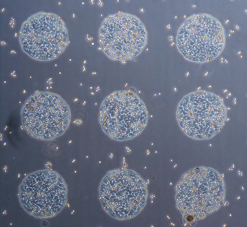
The application of defined amounts of cells at defined locations is an important topic in cellular biotechnology, e.g. in the field of tissue engineering, cell analysis and transfection arrays. The controlled release of living cells onto the corresponding substrate is crucial. With the technology available at the institute, living cells can be dispensed specifically onto surfaces to make them available for further investigations. The survival rate of the cells is up to 95 %. Thus, a technique is available to address a wide range of different questions. In transfection arrays the cells are dispensed onto a mixture of transfection reagent and DNA, which causes the cells to take up the DNA. The DNA can be protein-coding vectors or RNAi constructs, for example. After transfection, the cells can be characterized. With this method it is possible to test a large number of DNA constructs in parallel and under identical conditions. Another application of cell microarrays is the dispensing of different cell types onto (pre-)structured surfaces. This allows on the one hand to study and characterize the interaction of the cells and on the other hand to create tissue-like structures and cell populations.
Peptide Microarrays
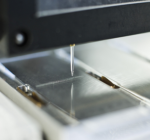
A challenge in protein microarrays is the functional application of membrane proteins. Peptide microarrays are an alternative. Here, proteins can be represented as peptides, from individual proteins to the entire proteome of cells. To produce peptide microarrays, high-density peptide microarrays are applied to SPOT synthesis or similar methods. At the institute, customized peptide microarrays are usually produced, where the peptides are purchased and after quality control immobilized in defined concentrations or series in multiple replicates. In contrast to proteins, peptides can differ greatly in their physicochemical properties such as isoelectric point and hydrophobicity. With the available technology, the ideal / optimal conditions can be specified for each individual peptide and all peptides can be dispensed reliably and reproducibly in very small volumes onto a wide variety of surfaces and structures.
Applications for peptide microarrays include the identification of antigenic regions of surface proteins in bacterial germs and the identification of immunogens, epitope mapping and determination of antibody specificity, serum profiling for the identification and validation of biomarkers, characterization of enzymes (e.g. target identification) or medical applications such as HLA profling and patient stratification.
Protein Microarrays
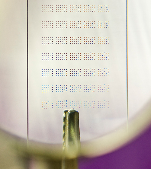
In protein microarrays, proteins are applied and immobilised on modified slides. For this purpose, the corresponding proteins can either be produced recombinantly, from cDNA or expression banks, or protein microarrays can be produced from DNA microarrays using in vitro transcription / translation on demand.
The produced protein microarrays can be used for many different applications. Most often these microarrays are used to identify interactions. Besides protein-protein interactions, interactions between immobilized proteins with small molecules, e.g. ATP and drugs, the influence of co-factors, salinity and pH on interactions, with lipids and carbohydrates can be investigated.
The challenge in production is to ensure that all proteins are immobilised uniformly and as functionally as possible. Numerous different techniques are available for this purpose. They range from adsorption on nitrocellulose, covalent coupling e.g. to epoxy-modified surfaces or by click chemistry, to immobilization via affinity tags or antibodies.
Due to the variety of available technologies and years of expertise, many protein classes can be applied to a wide variety of surfaces and made available for analysis.
DNA and RNA microarrays/expression analysis
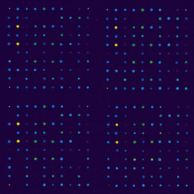
For the understanding of complex regulatory mechanisms and the study of cellular mechanisms, a simultaneous analysis of the expression of all transcripts of a cell, a cell population, a tissue or an organism during a defined period of time is indispensable. The complete genomic sequence of many organisms such as humans, mice, Drosophila melanogaster and Caenorhabditis elegans are now available. Using second-generation sequencing, information on the transcriptional activity of an organism can be determined within a few days. The method enables the identification of genes required for complex developmental networks and signal transduction pathways.
Based on this information, specific DNA / RNA microarrays can be produced to determine changes in selected significant transcripts. This information can be used, for example, to classify / stratify patients or to develop tests to support therapy decisions and assistance.
The focus is on the study of human cancer material. Information is collected to enable early diagnosis, accurate prognosis, identification of potentially interesting therapies and evaluation of the success of disease treatment. In addition to the analysis of RNA transcripts, microRNAs but also epigenetic changes can be determined.
Serum Screening
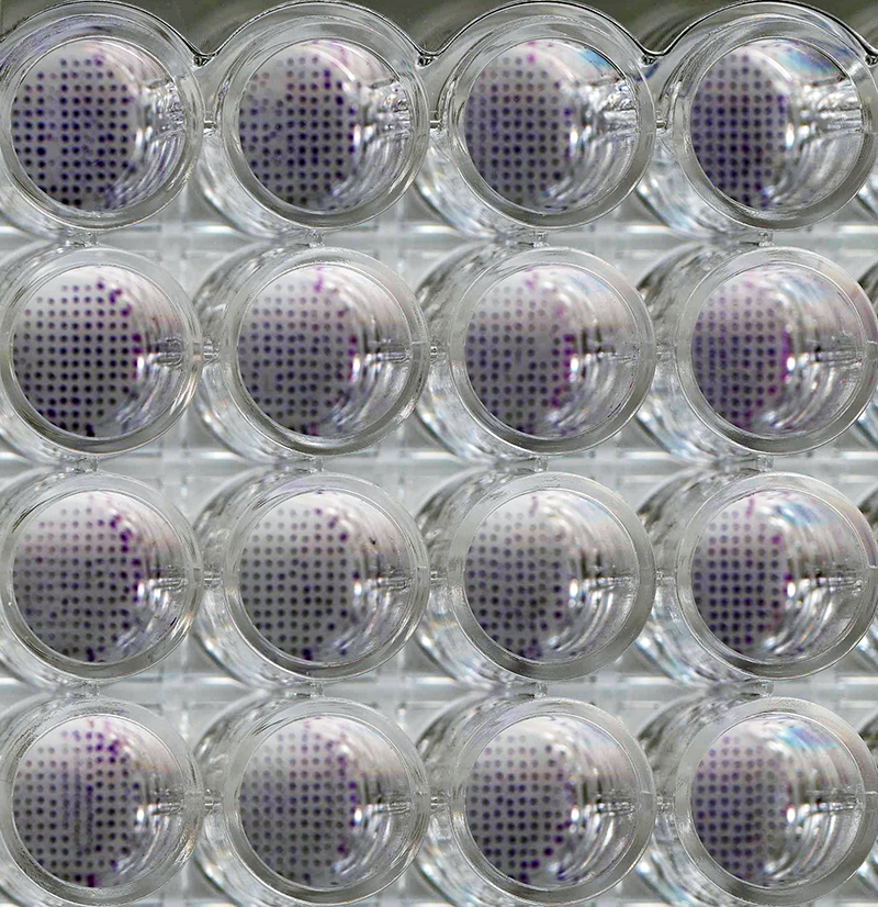
A major application of protein and peptide microarrays is the identification of antibodies in patient serum. This can be disease-associated antibodies, e.g. for cancer and rheumatism of the IgG and Ig M type, but also antibodies for allergies (type IgE) or against bacterial and viral infections. Depending on the problem, the proteins, parts of the proteins as peptides or the entire (sub-)proteome are immobilized on a microarray and incubated with serum. Detection is performed by means of specific antibodies against the corresponding IgG family. If necessary, the serum can be purified and the slide incubated with the concentration normalized IgGs. Besides a careful selection of potential antigens, the selection of patient sera is a critical factor. Selection here means a possible stratification of patients into subfamilies, the selection of the appropriate control patient cohort, the number of patients, the time of sampling(s) and the preparation and storage of the sera from sampling to transport and analysis. Based on these factors, microarrays can be a very powerful tool to identify and validate disease-associated biomarkers.
In addition, such microarrays can be used to determine serotypes and, based on the data obtained during transplantation, to make a prediction of the tolerance or risk of graft rejection. To date, well over 1,000 different HLA genotypes have been described. Only about 100 genotypes are available as proteins for analysis. The limitation here is the complex expression and purification of the HLA proteins, where the 3-dimensional conformation must be preserved. Protein microarrays or peptide microarrays with cyclized peptides can be a valuable tool to overcome these limitations.
Antibody Characterization
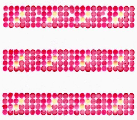
Well characterized and validated antibodies are essential for all microarray-based methods. The data obtained depend mainly on the quality of the antibodies used. Current studies show that in some cases less than 20% of the used and commercially available antibodies can be used for assay development and meaningful (clinical) studies. Due to cross-reactivity with other targets, non-specific antibodies can misrepresent the amount of antigen (yes/no response), lead to false positive and negative classifications and influence the biological / medical statement of an experiment.
In general, each reagent (antigen and antibody) should be subjected to a thorough quality control prior to an experiment. This includes new batches and batches of reagents for new orders.
Here the department has acquired extensive expertise due to years of work in the development of assays in the (bio)analytical field. In addition to classical methods such as ELISA and Western blot analyses, antibodies can be characterised and kinetic data obtained using peptide microarrays. Especially in the development of diagnostic and (bio)analytical methods, the type of antibody characterization is crucial. For example, antibodies can specifically recognize their target in one assay, but show cross reactivities on another platform under different conditions. The existing knowledge about the transferability of results between different platforms and techniques is an important expertise of the department in this field.
Analysis of signal paths
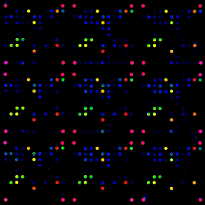
In addition to in vivo methods, a number of in vitro methods have become established in recent years for the analysis of signalling pathways. Reverse phase microarrays are one of the methods that are becoming more and more popular. Here the extract of cells e.g. from a biopsy or of cells with and without stimulus is spotted in a concentration series on slides and incubated with specific antibodies (e.g. phospho-specific antibodies). The signal intensity allows conclusions about changes in the proteins. Changes can be e.g. the abundance of the proteins or post-translational modifications. The advantages of microarrays are the small sample volume, a high number of technical replicates and the possibility to test a large number of samples under identical conditions. This allows the investigation of time courses as well as the analysis of signaling pathways or the "reaction" of cells (tissue) to external stimuli in order to draw conclusions about the mechanism of action.
Tabbed contents
Range of services
Range of services
- Production (spotting) of customized DNA, peptide, protein and cell microarrays
- Multiparameter analytics
- Competence Center Dispensing Technologies
- Spotting on different materials, e.g. glass, plastic, membranes, microtiter plates, conductor paths, etc.
- Benchmarking of various contact and non-contact spotters to select the optimal system (reference laboratory for liquid dispensing systems)
- Execution of customer-specific microarray experiments, evaluation and documentation
- Optimization of experiments by thermodynamic and kinetic measurements
- Transfer of other assay formats to microarrays, as lateral flow test and for in vitro diagnostic applications
- Development and establishment of assays for ELISA and microarrays
- Colorimetric, fluorescence and electrochemical detection systems
Methods and Devices
Methods
- Dispensing of biological fluids / samples in pL to µL volume
- Contact and non-contact based methods
- Dispensing on different surfaces
- Immobilization of (bio)molecules
- Quality control of the manufactured microarrays
- Surface Characterization
- Determination of the loading / immobilization density
- Modifications and coatings of surfaces
Devices
- I2-400 (Company M2)
- S5 (Company Scienion)
- S7 (Company Scienion)
- S100 (Company Scienion)
- Genetics
- Top spotter
- Contact angle measurement, ellipsometry
- bScreen (Biamterics)
 Fraunhofer Institute for Cell Therapy and Immunology, Branch Bioanalytics and Bioprocesses IZI-BB